Opthalmology
One of the most active fields of work in the VARPA Group is the ophtalmology, in particular the analysis of eye fundus images (retinal images).
The retinal image processing is a very interesting and demanding field, having a lot of practical applications, such as the development of applications for massive medical revision and the research in pharmacology effectiveness.
Collaborations
- Complexo Hospitalario Universitario de A Coruña
- Complexo Hospitalario Universitario de Santiago de Compostela
- Hospital de Conxo
- Hospital Naval de Ferrol
Working fields
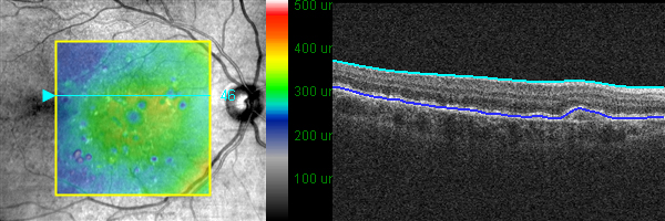
Drusen detection
Drusen are tiny white or yellow spots associated with the age-related macular degeneration (AMD). This disease can lead to severe loss central vision and adversely affect the patient's quality of life. We have developed an automatic methodology to detect drusen in initial stages. This methodology is based a template matching technique to find the drusen in the interest areas. Currently, our work is focused on the analysis of the drusen evolution in OCTR sequences.
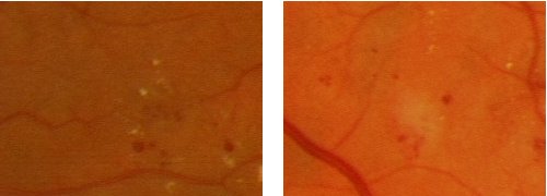
Red lesion detection
The diabetic retinopathy is a disease related to the diabetes that can cause blindness. One important symptom of diabetic retinopathy is the development of red lesions in the retina. We have developed an automatic methodology to analyse the retinal images in order to find this red lesions. This methodology is based on correlation filters and region growing segmentation techniques.
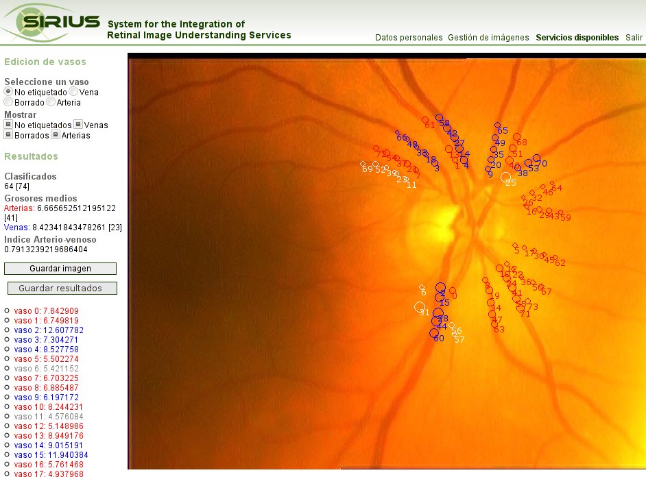
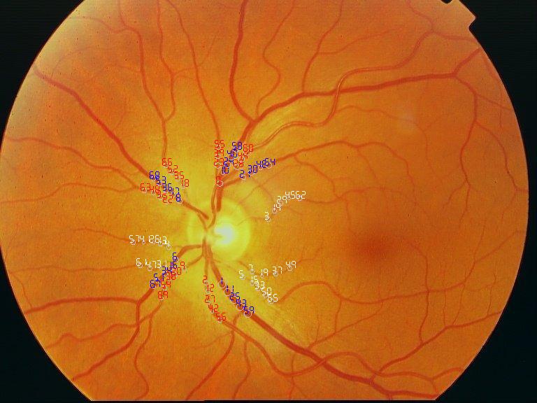
Arteriolar-to-Venular Ratio Computation
The retina Arteriolar-to-Venular ratio (AVR) is a parameter that helps the diagnosis of some pathologies, such as hypertension or arteriosclerosis. It is mainly computed as the ratio between the sum of artery calibers and the sum of vein calibers in several circumferences centered at the optic disc. We are currently developing and testing an automatic methodology to compute the AVR. This methodology involves the following steps:
- Optic disc location. It is the starting point. We are testing several algorithms to perform an efficient and reliable detection to the optic disc.
- Selection of Interest Radii centered at the optic disc.
- Vessel Detection and Caliber Measurement. We have developed a methodology based on snakes to measure the vessel diameter in concentric circumferences.
- Vessel Classification into Arteries and Veins. This is the most difficult step in the whole process due to the variability within inter-patient and intra-patient vessel contrast. We are testing several techniques and classifiers in order to minimize the misclassifications.
- AVR Computation. In the final step, we compute the AVR using the information obtained in the previous steps.
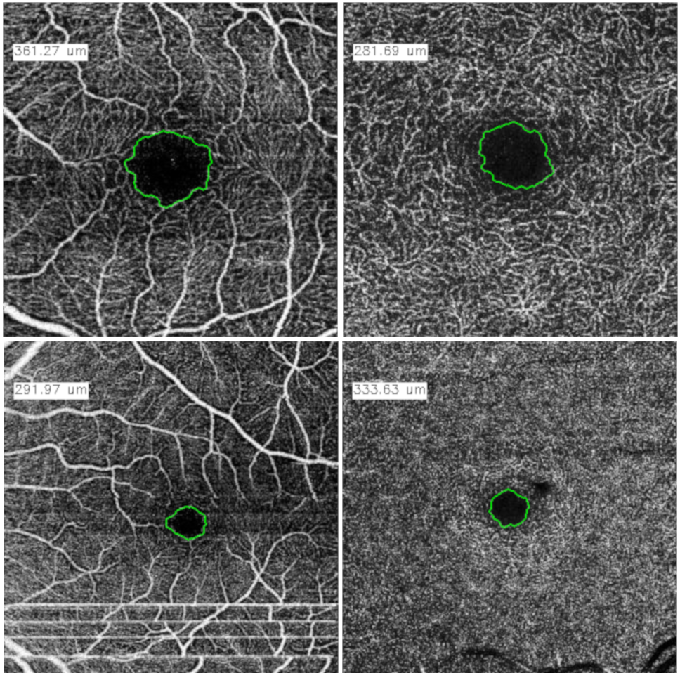
OCT-A
Angiography by Optical Coherence Tomography (OCT-A) is a non-invasive retinal imaging modality of recent appearance that allows the visualization of the vascular structure at predefined depths based on the detection of the blood movement through the retinal vasculature. In this way, OCT-A images allow us to measure the characteristics of the foveal vascular and avascular zones. Extracted parameters of this region can be used as prognostic factors that determine if the patient suffers from certain pathologies. The manual extraction of these biomedical parameters is a long, tedious and subjective process, introducing a significant intra and inter-expert variability, which penalizes the utility of the measurements. Our goals are the development of a fully automatic system that objectively segment and measure the region of the foveal avascular zone (FAZ) using this novel ophthalmological image modality, to ease the expert's work, increasing their productivity and making viable the use of this type of vascular biomarkers.
Applications
Sirius
Our goals are the development of user-friendly applications for support diagnosis and disease monitoring. In this sense, we are working in the development of a complete application for the automatic computation of the Arteriolar-to-Venular ratio, the SIRIUS web application. The use of web technologies makes easier the software maintenance and the collaboration of several experts from different medical institutions.
Public databases
VICAVR database
The VICAVR database is a set of retinal images used for the computation of the A/V Ratio. The database currently includes 58 images. The images have been acquired with a TopCon non-mydriatic camera NW-100 model and are optic disc centered with a resolution of 768x584. The database includes the caliber of the vessels measured at different radii from the optic disc as well as the vessel type (artery/vein) labelled by three experts.
If you are interested in using the VICAVR database, please send an email to: noelia.barreira@udc.es and you will receive an authentication password to access the database. This is intended for statistical purposes only, no private data or fee is required. If you use this image set for your work, please include a reference of VICAVR in it.
OCTAGON dataset
The OCTAGON dataset is a set of Angiography by Octical Coherence Tomography images (OCT-A) used to the segmentation of the Foveal Avascular Zone (FAZ). The dataset includes 144 healthy OCT-A images and 69 diabetic OCT-A images, divided into four groups, each one with 36 and about 17 OCT-A images, respectively. These groups are: 3x3 superficial, 3x3 deep, 6x6 superficial and 6x6 deep, where 3x3 and 6x6 are the zoom of the image and superficial/deep are the depth level of the extracted image. The healthy dataset includes OCT-A images from people classified in 6 age ranges: 10-19 years, 20-29 years, 30-39 years, 40-49 years, 50-59 years and 60-69 years. Each age range includes 3 different patients with information of left and right eyes for each one. Finally, for each eye, there are four different images: one 3x3 superficial image, one 3x3 deep image, one 6x6 superficial image and one 6x6 deep image. Each image have two manual labelled of expert clinicians of the FAZ and their quantification in the healthy OCT-A images, and one manual labelled in the diabetic OCT-A images.
If you are interested in using the OCTAGON database, please send an email to: joaquim.demoura@udc.es and you will receive an authentication password to access the database. This is intended for statistical purposes only, no private data or fee is required. If you use this image set for your work, please include a reference of OCTAGON in it.
FOCTAIR dataset
FOCTAIR: Fluorescein and Optical Coherence Tomography Angiography image registration dataset. This dataset contains a total of 86 cases obtained from a total of 29 patients. Each case corresponds to a patient's eye and has three images: one FA image (1536x1536 px) and two OCTA images of the superficial capillary plexus corresponding to two different zoom levels: 3x3mm and 6x6mm (both have the same size: 320x320 px.). In total, there are 172 possible registrations (86 pairs of FA and OCTA 3x3mm and 86 pairs of FA and OCTA 6x6mm).
If you are interested in using the FOCTAIR dataset, please send an email to: joaquim.demoura@udc.es and you will receive an authentication password to access the database. This is intended for statistical purposes only, no private data or fee is required. If you use this image set for your work, please include a reference of FOCTAIR in it.
CLOUD dataset
The CLOUD dataset is a set of Optical Coherence Tomography of the Anterior Segment images (AS-OCT) used to the automatic identification and representation of the cornea-contact lens relationship. The dataset includes 112 AS-OCT images that were captured from 16 different patients. In particular, the images were obtained by an OCT Cirrus 500 scanner model of Carl Zeiss Meditec with an anterior segment module for users of scleral contact lens (SCL).
If you are interested in using the CLOUD database, please send an email to: joaquim.demoura@udc.es and you will receive an authentication password to access the database. This is intended for statistical purposes only, no private data or fee is required. If you use this image set for your work, please include a reference of CLOUD in it.
- Cabaleiro, P.; de Moura, J.; Novo, J.; Charlón, P.; Ortega, M. "Automatic Identification and Representation of the Cornea–Contact Lens Relationship Using AS-OCT Images", Sensors, 19, 5087, 2019.
Main publications
-
,
,
,
,
,
,
"SIRIUS: A Web-based system for retinal image analysis",
International Journal of Medical Informatics,
79(10),
722-732,
2010.
[Abstract] [PDF] [+Info.]
-
,
,
,
,
,
"Improvements in Retinal Vessel Clustering Techniques:Towards the Automatic Computation of the Arterio Venous Ratio",
Computing, Archives for Scientific Computing,
90 (3),
197-217,
2010.
[Abstract] [PDF] [+Info.]
-
,
,
,
,
"The significance of the vessel registration for a reliable computation of arteriovenous ratio",
International Conference on Image Analysis and Recognition (ICIAR),
Aveiro, Portugal,
June 2012.
(pending of publication).
[Abstract] [PDF]
-
,
,
,
,
"Automatic extraction of vascularity measurements using OCT-A images",
Knowledge-Based and
Intelligent Information & Engineering Systems: Proceedings of the
22st International Conference, KES-2018,
126,
273-281,
Belgrado, Serbia,
September 2018.
[Abstract] [PDF]
-
,
,
,
,
,
,
"Automatic segmentation of the Foveal Avascular Zone in ophtalmological OCT-A images",
PLoS One,
14(2),
e0212364,
2019.
[Abstract]